
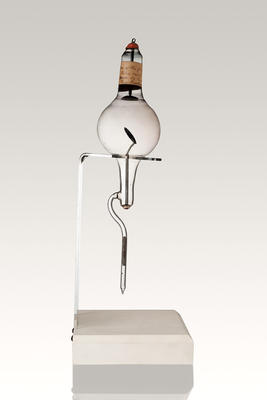
X-ray tube with internal cluster target
Maker
William Stone (b.1858, d.1949)
Date
Circa 1905
Description
X-ray tube with stone 'target' in claret and grey. Bulbous pear-shape with narrow cylindrical base. On turned wooden stand.
In 1895 Wilhelm Conrad Roentgen showed that for fifty years, scientists using Crookes tubes to study electricity had been unknowingly producing penetrating X-rays with the potential to make photographs of the living skeleton. Scientists were quick to apply this new information and researched ways to optimise the quality of X-rays emerging from the Cookes tubes in order to get better images.
In March 1896 Sir Thomas Ranken Lyle, professor of natural philosophy at the University of Melbourne, made a radiograph of the foot of his colleague Professor Orme Masson, probably the first of its kind in Australia. This X-ray tube is the work of William Stone, and Lyle and other early pioneers such as FJ Clendinnen made their own tubes as well as using those made by Stone. Stone was a friend of Lyle and was employed by the Victorian Railways, becoming chief electrical engineer in 1913. In 1903 he was appointed to the Faculty of Engineering at the University of Melbourne.
See full details


Do you know something about this object?
Be the first to comment on this object record.